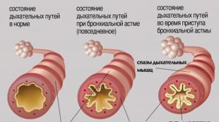
Discovery of viruses by Ivanovsky. I
The history of virology began at the end of the 19th century. after microbiology - the discoveries of L. Pasteur R. Koch and their employees. The discoverer of viruses was D. I. IVANOVSKY (1892), who showed that the causative agent of tobacco mosaic disease is able to pass through a filter that traps the smallest bacteria and does not grow on art, nutrient media.
Starting in 1887 to study tobacco diseases on the territory of Bessarabia and the Nikitsky Botanical Garden, he distinguished between the previously mixed so-called vendace and mosaic disease. He found out (1892) that the causative agent of the latter, unlike bacteria, is invisible in a microscope at the highest magnification, passes through porcelain filters and does not grow on ordinary nutrient media. He found crystalline inclusions (“Ivanovsky crystals”) in the cells of diseased plants, thus opening a special world of pathogens of non-bacterial and non-protozoal nature, later called viruses. Ivanovsky considered them as the smallest living organisms. In addition, Ivanovsky published works on the features of physiological processes in diseased plants, the effect of oxygen on alcoholic fermentation in yeast, the state of chlorophyll in plants, its resistance to light, the importance of carotene and xanthophyll, and on soil microbiology.
The main results of observations and studies of the anatomy and physiology of diseased plants (“On Diseases of Tobacco Plants” - Proceedings of the St. Petersburg Society of Naturalists, vol. 19) were reported by D. I. Ivanovsky in 1888 at a meeting of the St. Petersburg Society of Naturalists and are presented in the article D. I. Ivanovsky and V. V. Polovtsev, and also published in the Proceedings of the Imperial Free Economic Society in 1889, and then in the brochure “Ryabukha is a disease of tobacco, its causes, means of combating it” (St. Petersburg, 1890) in the same year, republished in German by the Russian Academy of Sciences. As a result of these observations, D.I. Ivanovsky and V.V. Polovtsov was the first to suggest that the tobacco disease, described in 1886 by A. Mayer in Holland under the name mosaic, represents not one, but two completely different diseases of the same plant; one of them is vendace, the causative agent of which is a fungus, and the other is of unknown origin. Based on the experience of the peasants, their own observations and the study of diseased plants, D.I. Ivanovsky and V.V. Polovtsev came to the conclusion that the venison disease affects plants planted on old tobacco plantations, and gave recommendations on the introduction of crop rotation and improving the culture of farming. and means of dealing with it.
D.I. Ivanovsky showed that the disease of tobacco - tobacco mosaic - can be transferred from diseased plants to healthy ones if they are infected with the juice of diseased plants, previously passed through a special filter that traps bacteria. The causative agent of mosaic disease is called by D.I. Ivanovsky either “filtering bacteria”, or microorganisms, and this is understandable, since it was very difficult to immediately formulate the existence of a special world of viruses. In 1898, M. Beijerink confirmed the data of D.I. Ivanovsky and put forward the hypothesis that the disease is not caused by a bacterium, but by a fundamentally new, different from bacteria, infectious agent. He called it contagium vivum fluidum (liquid infectious principle), in other words, a filterable virus (the term “virus” - from the Latin “poison”, “poisonous principle” - was then used to refer to the infectious beginning of any disease).
In addition to capital conclusions confirming the existence of a new, previously unknown class of microorganisms, Ivanovsky's dissertation contains other important data. This is how he described the cytopathic action of the causative agent of tobacco mosaic and gave a characteristic of the crystals, which later, in 1935, were identified as crystals of the tobacco mosaic virus. There is also a description of intracellular inclusions, which laid the foundation for the study of inclusions in viral infections, which has retained its significance for the diagnosis of viral diseases.
Along with the work on virology, which brought him worldwide fame, he also conducted other studies. He has 180 publications, including a number of works in the field of soil microbiology, plant physiology and anatomy, and 30 articles in the Brockhaus Encyclopedic Dictionary. Ivanovsky's scientific activity was combined with pedagogical activity: he was an excellent lecturer and teacher who brought up more than one generation of students at St. Petersburg, Warsaw and Donskoy universities.
ETIOLOGY - the doctrine of the causes and conditions for the occurrence and development of diseases and pathological processes.
ETIOLOGICAL FACTOR (EF) - the main, leading, causing factor, without the presence of which there would be no disease (for example, Koch's bacillus in tuberculosis). The etiological factor can be simple (mechanical effect) or complex (damaging factors of a nuclear explosion), acting for a long time, throughout the entire disease (microbes, viruses, toxins), or only triggering a pathological process (thermal factor during a burn).
DISEASE PATHOGENESIS is a dialectically contradictory process that includes two opposite tendencies: on the one hand, these are the mechanisms of sex, damage, deviation from the norm, and on the other hand, the mechanisms of protection, adaptation, compensation and reparation.
Examples, HPV are small DNA viruses, the characteristic feature of which is the ability to cause proliferation of the epithelium of the skin and mucous membranes.
Back in 1986 Ya.V. Bohman formulated general principles for the treatment of background diseases of the cervix: it should include the elimination of the etiological factor and those inflammatory, dyshormonal, immunosuppressive and dysmetabolic changes in the body that contribute to its occurrence and maintenance of a long course.
Currently, etiopathogenetic therapy of cervical pathology has two main directions - the impact on the etiological factor - human papillomavirus and the blocking of the main mechanisms of carcinogenesis, namely hormonal carcinogenesis associated with increased formation of the aggressive metabolite of estradiol - 16a-hydroxyestrone (16a-OH) against the background of hyperestrogenemia and HPV infection.
The etiological role of human herpesvirus type 6 in human pathology has been proven in relation to two independent diseases: sudden exanthema in young children and chronic fatigue syndrome in adults.
Later, the etiological role of HHV-6 in the occurrence of this syndrome was proven. Although it remains unclear to date whether chronic fatigue syndrome is etiologically associated with infection with the human herpesvirus type 6, or whether the disease is a consequence of the reactivation of a latent infection, i.e. HHV-6 plays a pathogenetic role. Unlike sudden exanthema, chronic fatigue syndrome is a disease of adults.
In 1957, polio, Coxsackie and ECHO viruses were grouped together and named human enteric viruses. Over the past decades, facts have continued to accumulate explaining the role of enteroviruses in human pathology.
As has now been established, enteroviruses play an etiological role in the occurrence of serous meningitis and, much less frequently, meningoencephalitis with a benign course.
Discovery of viruses
Virology is a young science with a history of just over 100 years. Having begun its journey as the science of viruses that cause diseases in humans, animals and plants, virology is currently developing in the direction of studying the basic laws of modern biology at the molecular level, based on the fact that viruses are part of the biosphere and an important factor in the evolution of the organic world.
History of virology is unusual in that one of its subjects, viral diseases, began to be studied long before the actual viruses were discovered. The beginning of the history of virology is the fight against infectious diseases, and only subsequently - the gradual disclosure of the sources of these diseases. This is confirmed by the work of Edward Jenner (1749-1823) on the prevention of smallpox and the work of Louis Pasteur (1822-1895) with the causative agent of rabies.
Since time immemorial, smallpox has been the scourge of mankind, claiming thousands of lives. Descriptions of smallpox infection are found in the manuscripts of the most ancient Chinese and Indian texts. The first mention of smallpox epidemics on the European continent dates back to the 6th century AD (an epidemic among the soldiers of the Ethiopian army besieging Mecca), after which there was an inexplicable period of time when there was no mention of smallpox epidemics. Smallpox began to roam the continents again in the 17th century. For example, in North America (1617-1619), 9/10 of the population died in Massachusetts, in Iceland (1707) after a smallpox epidemic, only 17 thousand people remained from 57 thousand people, in the city of Eastham (1763 ) out of 1331 inhabitants, 4 people remained. In this regard, the problem of combating smallpox was very acute.
The method of preventing smallpox through vaccination, called variolation, has been known since ancient times. References to the use of variolation in Europe date back to the middle of the 17th century with references to earlier experience in China, the Far East, and Turkey. The essence of variolation was that the contents of pustules from patients who had a mild form of smallpox were introduced into a small wound on the human skin, which caused a mild illness and prevented an acute form. However, at the same time, a great danger of a severe form of smallpox remained, and mortality among the vaccinated reached 10%. Jenner revolutionized smallpox prevention. He was the first to draw attention to the fact that people who had had cowpox, which proceeded easily, subsequently never had smallpox. On May 14, 1796, Jenner introduced into the wound of James Phips, who had never had smallpox, liquid from the pustules of Sarah Selmes, a milkmaid who had cowpox. At the site of artificial infection, the boy developed typical pustules, which disappeared after 14 days. Then Jenner introduced a highly infectious material from the pustules of a smallpox patient into the boy's wound. The boy didn't get sick. This is how the idea of vaccination was born and confirmed (from the Latin word vacca - a cow).
In Jenner's time, vaccination was understood as the introduction of vaccinia infectious material into the human body with the aim of preventing smallpox from contracting smallpox. The term vaccine was applied to a substance that prevented smallpox. Since 1840, smallpox vaccine began to be obtained by infecting calves. The human smallpox virus was discovered only in 1904. Thus, smallpox is the first infection against which a vaccine was used, that is, the first controllable infection. Advances in vaccination against smallpox have led to its eradication on a global scale.Nowadays, vaccination and vaccine are used as general terms for inoculation and inoculation material.
Pasteur, who essentially did not know anything concrete about the causes of rabies, except for the indisputable fact of its infectious nature, used the principle of weakening (attenuation) of the pathogen. In order to weaken the pathogenic properties of the rabies pathogen, a rabbit was used, into the brain of which the brain tissue of a dog that had died of rabies was injected. After the death of a rabbit, its brain tissue was introduced to the next rabbit, and so on. About 100 passages were carried out before the pathogen adapted to the brain tissue of the rabbit. Being introduced subcutaneously into the body of a dog, it showed only moderate properties of pathogenicity. Such a "re-educated" pathogen Pasteur called "fixed", in contrast to the "wild", which is characterized by high pathogenicity. Later, Pasteur developed a method for creating immunity, consisting of a series of injections with a gradually increasing content of a fixed pathogen. The dog that completed the full course of injections was found to be fully resistant to infection. Pasteur came to the conclusion that the process of development of an infectious disease, in essence, is the struggle of microbes with the body's defenses. “Each disease must have its pathogen, and we must contribute to the development of immunity to this disease in the patient’s body,” said Pasteur. Still not understanding how the body develops immunity, Pasteur managed to use his principles and direct the mechanisms of this process to the benefit of man. In July 1885, Pasteur had the opportunity to test the properties of a "fixed" rabies agent on a child bitten by a rabid dog.
The boy was given a series of injections of an increasingly poisonous substance, with the last injection already containing a completely pathogenic form of the pathogen. The boy remained healthy. The rabies virus was discovered by Remlenge in 1903.It should be noted that neither the smallpox virus nor the rabies virus was the first discovered virus to infect animals and humans. The first place rightfully belongs to the foot-and-mouth disease virus, discovered by Leffler and Frosch in 1898. These researchers, using multiple dilutions of a filtering agent, showed its toxicity and made a conclusion about its corpuscular nature.
By the end of the 19th century, it became clear that a number of human diseases, such as rabies, smallpox, influenza, yellow fever, are infectious, but their pathogens were not detected by bacteriological methods. Thanks to the work of Robert Koch (1843-1910), who pioneered the pure bacterial culture technique, it became possible to distinguish between bacterial and non-bacterial diseases. In 1890, at the X Congress of Hygienists, Koch was forced to declare that "... in the listed diseases, we are not dealing with bacteria, but with organized pathogens that belong to a completely different group of microorganisms." This statement by Koch shows that the discovery of viruses was not a random event. Not only the experience of working with pathogens that are incomprehensible in nature, but also an understanding of the essence of what is happening contributed to the fact that the idea was formulated about the existence of an original group of pathogens of infectious diseases of a non-bacterial nature. It remained to experimentally prove its existence.
The first experimental proof of the existence of a new group of pathogens of infectious diseases was obtained by our compatriot, plant physiologist Dmitry Iosifovich Ivanovsky (1864-1920), while studying tobacco mosaic diseases. This is not surprising, since infectious diseases of an epidemic nature have often been observed in plants. Back in 1883-84. Dutch botanist and geneticist de Vries observed an epidemic of greening flowers and suggested the infectious nature of the disease. In 1886, the German scientist Mayer, who worked in Holland, showed that the sap of plants suffering from mosaic disease, when inoculated, causes the same disease in plants. Mayer was sure that the culprit of the disease was a microorganism, and unsuccessfully searched for it. In the 19th century, tobacco diseases caused great harm to agriculture in our country as well. In this regard, a group of researchers was sent to Ukraine to study tobacco diseases, which, being a student of St. Petersburg University, included D.I. Ivanovsky. As a result of studying the disease, described in 1886 by Mayer as a mosaic disease of tobacco, D.I. Ivanovsky and V.V.
Polovtsev came to the conclusion that it represents two different diseases. One of them - "ribbon" - is caused by a fungus, and the other is of unknown origin. The study of tobacco mosaic disease was continued by Ivanovsky in the Nikitsky Botanical Garden under the guidance of Academician A.S. Famycin. Using the juice of a diseased tobacco leaf, filtered through a Chamberlain candle, which retains the smallest bacteria, Ivanovsky caused a disease in tobacco leaves. Cultivation of infected juice on artificial nutrient media did not give results, and Ivanovsky comes to the conclusion that the causative agent of the disease has an unusual nature - it is filtered through bacterial filters and is not able to grow on artificial nutrient media. Heating the juice at a temperature from 60 °C to 70 °C deprived it of infectivity, which indicated the living nature of the pathogen. Ivanovsky first named the new type of pathogen "filterable bacteria" (Figure 1). The results of the work of D.I. Ivanovsky were the basis of his dissertation, presented in 1888, and published in the book "On Two Diseases of Tobacco" in 1892. This year is considered the year of the discovery of viruses.A - Electron micrograph after oblique deposition with carbon and platinum; 65,000 ´. (Photo by N. Frank.) B - Model. (Karlson, Kurzes Lehrbuch der Biochemie, Stuttgart, Thieme, 1980).
Figure 1 - Tobacco mosaic virus
For a certain period of time in foreign publications, the discovery of viruses was associated with the name of the Dutch scientist Beyerink (1851-1931), who also studied tobacco mosaic disease and published his experiments in 1898. Beyerink placed the filtered juice of an infected plant on the surface of an agar, incubated and obtained bacterial colonies on its surface. After that, the upper layer of agar with colonies of bacteria was removed, and the inner layer was used to infect a healthy plant. The plant is sick. From this, Beijerinck concluded that the cause of the disease was not bacteria, but some liquid substance that could penetrate the agar, and called the pathogen "liquid live contagion." Due to the fact that Ivanovsky only described his experiments in detail, but did not pay due attention to the non-bacterial nature of the pathogen, there was a misunderstanding of the situation. Ivanovsky's work gained fame only after Beijerinck repeated and expanded his experiments and emphasized that Ivanovsky proved for the first time precisely the non-bacterial nature of the causative agent of the most typical viral disease of tobacco. Beijerinck himself recognized the primacy of Ivanovsky and, at present, the priority of the discovery of viruses by D.I. Ivanovsky is recognized all over the world.
Word VIRUS means poison. This term was used by Pasteur to refer to the contagious beginning. It should be noted that in the early 19th century, all pathogenic agents were called the word virus. Only after the nature of bacteria, poisons and toxins became clear, the terms "ultravirus", and then simply "virus" began to mean "a new type of filterable pathogen." The term “virus” took root widely in the 30s of our century.
It is now clear that viruses are characterized by ubiquitousness, that is, the ubiquity of distribution. Viruses infect representatives of all living kingdoms: humans, vertebrates and invertebrates, plants, fungi, bacteria.
The first report relating to bacterial viruses was made by Hankin in 1896. In the Chronicle of the Pasteur Institute, he stated that "... the water of some rivers of India has a bactericidal effect ...", which is undoubtedly associated with bacterial viruses. In 1915, Twoorth in London, studying the causes of lysis of bacterial colonies, described the principle of transmission of "lysis" to new cultures in a series of generations. His work, as often happens, actually went unnoticed, and two years later, in 1917, the Canadian de Hérelle rediscovered the phenomenon of bacterial lysis associated with a filtering agent. He named this agent bacteriophage. De Hérelle assumed that there was only one bacteriophage. However, studies by Barnet, who worked in Melbourne in 1924-34, showed a wide variety of bacterial viruses in physical and biological properties. The discovery of the diversity of bacteriophages aroused great scientific interest. At the end of the 1930s, three researchers - the physicist Delbrück, the bacteriologists Luria and Hershey, who worked in the USA, created the so-called "Phage Group", whose research in the field of bacteriophage genetics ultimately led to the birth of a new science - molecular biology.
The study of insect viruses has lagged far behind the virology of vertebrates and humans. It is now clear that insect-infecting viruses can be conditionally divided into 3 groups: insect viruses themselves, animal and human viruses, for which insects are intermediate hosts, and plant viruses, which also infect insects.
The first insect virus to be identified is the silkworm jaundice virus (silkworm polyhedrosis virus, named Bollea stilpotiae). As early as 1907, Provacek showed that the filtered homogenate of diseased larvae is infectious for healthy silkworm larvae, but it was not until 1947 that the German scientist Bergold discovered rod-shaped viral particles.
One of the most fruitful studies in the field of virology is Reid's study of the nature of yellow fever on US Army volunteers in 1900-1901. It has been convincingly demonstrated that yellow fever is caused by a filterable virus transmitted by mosquitoes and mosquitoes. Mosquitoes were also found to remain non-infectious after ingesting infectious blood for two weeks. Thus, the external incubation period of the disease (the time required for the reproduction of the virus in the insect) was determined and the basic principles of the epidemiology of arbovirus infections (viral infections transmitted by blood-sucking arthropods) were established.
The ability of plant viruses to reproduce in their vector, the insect, was shown in 1952 to Maramoros. The researcher, using an insect injection technique, convincingly demonstrated the ability of the aster jaundice virus to multiply in its vector, the six-spotted cicada.
BIOLOGY
ON THE TOPIC:
"VIRUS"
Pupil 9B class
Valdai
Novgorod region
Shakhov Vladimir
Vladimirovich
Teacher: Ignatieva
Tat `yana Aleksandrovna
Introduction
a). First meeting;
b). Components of viruses;
in). Lysogeny;
G). Discovery of Hershey and Chase;
ΙΙΙ. Virus commandments.
Ι V. How are viruses arranged?
a). The nature of the virus;
b). Viruses are the kingdom of living organisms;
G). chemical composition of viruses;
V. Who are their parents?
VΙ. Interaction of a virus with a cell.
VΙΙ. Classification of viruses.
VΙΙΙ. The role of viruses in human life. Methods of transmission of viral diseases.
Ι X. List of black cases of viruses:
a). Flu;
b). smallpox;
in). Polio;
G). Rabies;
e). Viral hepatitis;
e). tumor viruses;
and). AIDS.
X. Statistical data on viral diseases and vaccinations (vaccination) according to MOSSh No. 2 in Valdai
XΙ. Features of the evolution of viruses at the present stage.
Conclusion.
Bibliography.
Introduction.
About the kingdoms that we see and do not see.
The fabulous concept of "kingdom" has taken root in science. There is the kingdom of plants, animals and the invisible kingdom of viruses. The first two kingdoms coexist relatively peacefully with each other, and the third invisible is aggressive and insidious. Its representatives do not like to live in peace either with each other or with others. Viruses live while they fight and die from inactivity. They are very whimsical about food, they live "on loan" at the expense of animal cells, plants and even bacteria. Viruses bring mostly harm and very rarely benefit, so to speak, benefit through harm.
The kingdom of viruses was discovered relatively recently: 100 years is a child's age compared to mathematics, 100 years is a long time compared to genetic engineering. Science has no age: science, like people, has youth, science is never old.
In 1892, the Russian scientist D.I. Ivanovsky described the unusual properties of the causative agents of tobacco disease - (tobacco mosaic), which passed through bacterial filters.
A few years later, F. Leffler and P. Frosch found that the causative agent of foot-and-mouth disease (a disease of livestock) also passes through bacterial filters. And in 1917, F. d'Errell discovered a bacteriophage - a virus that infects bacteria. So the viruses of plants, animals and microorganisms were discovered.
These three events marked the beginning of a new science - virology, which studies non-cellular life forms.
Viruses, although very small, they cannot be seen, are the object of study of the sciences:
For physicians, viruses are the most common causative agents of infectious diseases: influenza, measles, smallpox, tropical fevers.
For a pathologist, viruses are the etiological agents (cause) of cancer and leukemia, the most frequent and dangerous pathological processes.
For a veterinary worker, viruses are the culprits of epizootics (mass diseases) of foot-and-mouth disease, bird plague, infectious anemia and other diseases that affect farm animals.
For an agronomist, viruses are the causative agents of spotted banding of wheat, tobacco mosaic, yellow potato dwarfism and other diseases of agricultural plants.
For the grower, viruses are the factors that cause the amazing colors of tulips to appear.
For a medical microbiologist, viruses are agents that cause the appearance of toxic (poisonous) varieties of diphtheria or other bacteria, or factors that contribute to the development of bacteria resistant to antibiotics.
For an industrial microbiologist, viruses are pests of bacteria, producers, antibiotics, and enzymes.
For a geneticist, viruses are carriers of genetic information.
For a Darwinist, viruses are important factors in the evolution of the organic world.
For an ecologist, viruses are factors involved in the formation of conjugated systems of the organic world.
For a biologist, viruses are the simplest forms of life, possessing all of its main manifestations.
For the philosopher, viruses are the clearest illustration of the dialectics of nature, a touchstone for polishing such concepts as living and nonliving, part and whole, form and function.
Three main circumstances determined the development of modern virology, making it a kind of point (or kidney) of the growth of biomedical sciences.
Viruses are the causative agents of the most important diseases of humans, farm animals and plants, and their importance is constantly increasing as the incidence of bacterial, protozoal and fungal diseases decreases.
It is now recognized that viruses are the causative agents of cancer, leukemia and other malignant tumors. Therefore, the solution of the problems of oncology now depends on the knowledge of the nature of cancer pathogens and the mechanisms of carcinogenic (tumor-causing) transformations of normal cells.
Viruses are the simplest forms of life, possessing its main manifestations, a kind of abstraction of life, and therefore serve as the most grateful object of biology in general and molecular biology in particular.
Viruses are ubiquitous and can be found anywhere there is life. You can even say that viruses are a kind of "indicators of life." They are our constant companions and from the day of birth they accompany us always and everywhere. The damage they cause is very great. Suffice it to say that more than half of all human diseases are “on the conscience”, and if we remember that these smallest of the smallest ones still affect animals, plants and even their closest relatives in the microcosm - bacteria, it becomes clear that the fight against viruses is one of the top priorities. tasks. But in order to successfully fight the insidious invisibles, it is necessary to study their properties in detail.
Ι. Hypotheses of the origin of viruses.
Three main hypotheses have been put forward.
The possibility of degenerative evolution has been repeatedly established and proven, and perhaps the most striking example of it is the origin of some eukaryotic cell organelles from symbiotic bacteria. At present, based on the study of the homology of nucleic acids, it can be considered established that the chloroplasts of protozoa and plants originate from the ancestors of the current blue-green bacteria, and mitochondria from the ancestors of purple bacteria. The possibility of the origin of centrioles from prokaryotic symbionts is also discussed. Therefore, such a possibility is not excluded for the origin of viruses, especially such large, complex and autonomous ones as the smallpox virus is.
Nevertheless, the world of viruses is too diverse to admit the possibility of such a deep degenerative evolution for most of its representatives, from smallpox, herpes and iridoviruses to adenosatellites, from reoviruses to satellites of the tobacco necrosis virus or the RNA-containing delta virus - the satellite of the hepatitis virus AT, not to mention such autonomous genetic structures as plasmids or viroids. The diversity of genetic material in viruses is one of the arguments in favor of the origin of viruses from precellular forms. Indeed, the genetic material of viruses "exhausts" all its possible forms: single- and double-stranded RNA and DNA, their linear, circular and fragmentary types. Nature, as it were, tried all possible variants of genetic material on viruses before finally choosing its canonical forms - double-stranded DNA as the keeper of genetic information and single-stranded RNA as its transmitter. And yet, the diversity of the genetic material in viruses is more likely to indicate a polyphyletic origin of viruses than to the preservation of ancestral precellular forms, the genome of which evolved along an unlikely path from RNA to DNA, from single-stranded forms to double-stranded forms, etc.
The third hypothesis of 20-30 years seemed unlikely and even received the ironic name of the frenzied gene hypothesis. However, the accumulated facts give more and more arguments in favor of this hypothesis. A number of these facts will be discussed in a special part of the book. Here we note that it is this hypothesis that easily explains not only the quite obvious polyphyletic origin of viruses, but also the commonality of such diverse structures as complete and defective viruses, satellites and plasmids. It also follows from this concept that the formation of viruses was not a one-time event, but occurred many times and continues to occur at the present time. Already in ancient times, when cellular forms began to form, non-cellular forms, represented by viruses, autonomous, but cell-dependent genetic structures, were preserved and developed along with them. The currently existing viruses are products of evolution, both of their most ancient ancestors and of recently emerged autonomous genetic structures. Probably, tailed phages are an example of the former, while R-plasmids are an example of the latter.
ΙΙ. The history of the discovery of viruses.
First meeting.
In the 80s of the century in the south of Russia, tobacco plantations were subjected to a formidable invasion. The tops of plants died off, light spots appeared on the leaves, the number of affected fields increased from year to year, and the cause of the diseases is unknown.
Professors of St. Petersburg University, the world famous A. N. Beketov and A. S. Felintsin sent a small expedition to Bessarabia and Ukraine in the hope of understanding the causes of the disease. The expedition included D.I. Ivanovsky and V.V. Polovtsev.
DI. Ivanovo Russian scientist in 1892 discovered the tobacco mosaic virus.
Ivanovsky spent several years looking for pathogens. He collected facts, made observations, asked the peasants about the symptoms of the disease. And experimented. He collected leaves from several diseased plants. After 15 days, whitish spots appeared on these leaves. This means that the disease is indeed contagious, and can be transmitted from plant to plant. Ivanovsky consistently eliminated possible carriers of the disease - the root system of plants, seeds, flowers, pollen ... Experiments showed that they were not the point: the pathogenic principle infects plants in a different way.
Then the young scientist sets up a simple experiment. He collects diseased leaves, crushes them and buries them in areas with healthy plants. After a while, the plants get sick. So, the first success - the path from a diseased plant to a healthy one was found. The pathogen is transmitted by leaves that have fallen into the soil, overwinter and infect crops in spring.
But he did not know anything about the causative agent itself. His experiments showed only one thing - something contagious is contained in the juice. During these years, several more scientists in the world struggled to identify this “something”. A. Mayer in Holland suggested that the infectious principle is bacteria.
However, Ivanovsky proved that Mayer was mistaken in considering bacteria as carriers of the disease.
After filtering the infectious juice through fine-pored porcelain filters, he deposited bacteria on them. Now the bacteria have been removed... but the contagiousness of the juice has been preserved.
Six years pass and Ivanovsky discovers that he has encountered an incomprehensible agent that causes the disease: it does not multiply on artificial media, penetrates through the thinnest pores, and dies when heated. Filterable poison! This was the conclusion of the scientist.
But poison is a substance, and the causative agent of tobacco was a being. It reproduced well in the leaves of plants.
So Ivanovsky discovered a new realm of living organisms, the smallest of all living organisms and therefore invisible in a light microscope. Passing through the finest filters, remaining in the juice for years and at the same time not losing virulence. In 1889, the Danish botanist Martin Willem Beyrink, who became interested in the disease of tobacco Mayer, called the newly discovered creature a virus, adding that the virus is a "liquid, living, infectious principle."
Components of a virus
In 1932, the then director of the Rockefeller Institute in New York, Simon Fleckener, suggested that the young American biochemist Wendill Stanley study viruses. Stanley started by collecting a ton of tobacco leaves infected with tobacco mosaic virus and decided to extract the juice from the whole mountain. He squeezed out a bottle of juice and began to examine the juice with the chemical methods available to him. He exposed various fractions of juice to various reagents, hoping to get a pure viral protein (Stanley was convinced that a virus is a protein). For a long time he could not get rid of the proteins of plant cells. Once, having tried various methods of acidification and salting out, Stanley received an almost pure fraction of a protein that differed in its composition from the proteins of plant cells. The scientist realized that before him was what he so stubbornly sought. Stanley isolated an extraordinary protein, dissolved it in water and put the solution in the refrigerator. The next morning in the flask, instead of a clear liquid, there were beautiful silky needle-like crystals. Stanley extracted a tablespoon of such crystals from a ton of leaves. Then Stanley poured out some crystals, dissolved them in water, moistened gauze with this water and rubbed the leaves of healthy plants with it. Plant sap has undergone a whole range of chemical influences. After such a “massive processing”, the viruses, most likely, should have died.
The grated leaves got sick, and after a couple of weeks, a characteristic mosaic of white spots covered all the plants, then repeated this operation again, and after the fourth or fifth “transfusion” of the virus, he squeezed the juice from the leaves, subjected it to the same chemical treatment and again received exactly the same crystals. The strange properties of the virus were replenished with one more thing - the ability to crystallize.
The effect of crystallization was so overwhelming that Stanley for a long time abandoned the idea that the virus is a creature. Since all enzymes (catalysts for reactions in living organisms) are proteins, and many enzymes also increase in number as an organism develops and can crystallize, Stanley concluded that viruses are pure proteins, rather enzymes.
Soon, scientists became convinced that it was possible to crystallize not only the tobacco mosaic virus, but also a number of other viruses.
Wendel Stanley was awarded the Nobel Prize in 1946.
Five years later, the English biochemists F. Bowden and N. Piri found an error in Stanley's definition. 94% of the contents of the tobacco mosaic virus consisted of protein, and 6% was nucleic acid. The virus was not actually a protein, but a nucleoprotein - a combination of protein and nucleic acid.
As soon as electron microscopes became available to biologists, scientists found that virus crystals consist of several hundred billion particles tightly pressed together. There are so many particles in one crystal of the polio virus that they can infect all the inhabitants of the Earth more than once. When it was possible to examine individual viral particles in an electron microscope, it turned out that they come in different shapes - both spherical and rod-shaped, and in the form of a sandwich, and in the form of a mace, but always the outer shell of viruses consists of protein, and the internal content is represented by nucleic acid .
Lysogeny
When virologists got to know the life of viruses more closely, they discovered another unexpected property in them. Previously, it was believed that any particle of the virus, once it enters the cell, begins to multiply there and, in the end, the cell dies. But in 1921, and then in the mid-30s. years at the Pasteur Institute in Paris, a strange picture was described. Bacteriophages were added to the bacteria. After a certain period of time, the cells should have died, but, surprisingly, some of them remained alive and continued to multiply, despite the fact that phages were swarming with phages. Somehow, these cells became immune to the phages. Scientists isolated such cells, purified them of phages, then began to seed them regularly and one day found that in a phage-free culture of bacteria, out of nowhere, phage particles reappear.
Having disappeared for a while, as if hiding inside the cell, the phages again declared their existence. The same phages were tested on fresh, yet uninfected bacterial cultures. The phages were still behaving unusually. Some of them, as expected, caused cell death, but many disappeared inside the cells, and as soon as this happened, the cells gained the ability to resist infection by other similar viruses.
The process of the disappearance of viruses was called lysogenization, and cells infected with such viruses were called lysogenic. All attempts to detect all sorts of phages inside lysogenic bacteria ended in failure. The virus was attached to some structure of the cell and without it did not multiply.
Using a micromanipulator, scientists Lvov and Tutman separated one cell from the total mass of lysogenic bacteria and began to observe it. The cell divided once, giving rise to two young cells, which, in turn, gave rise to offspring after the allotted time. The cell suspected of hiding a bacterial virus inside was no different. Fifteen generations of bacteria changed, but patient scientists constantly observed with a microscope, replacing each other at regular intervals. During the 19th division, one of the cells burst, just like normal bacteria infected with a normal virus would burst.
Scientists have determined that lysogenic cells, although they carry the virus or part of it, but for the time being, this virus is not infectious. They called such an intracellular virus a provirus, or, in the case of bacteriophages, a prophage.
Then they proved that the provirus, once it enters the bacterium, does not disappear. After 18 generations, it was discovered. It remained to assume that all this time the prophage multiplied along with the bacterium.
Subsequently, it was proved that usually prophages cannot reproduce on their own, as all other viruses do, but multiply only when the bacterium itself reproduces.
And, finally, the third honor of this discovery belongs to Lvov, Siminovich and Kildgard - a method for isolating a provirus from a state of equilibrium. Influencing small doses of ultraviolet rays on lysogenic cells, it was possible to restore their ability to prophages to multiply independently of cells. Such released phages behaved exactly as their ancestors did: multiply and destroy cells. Lvov made the correct, only conclusion from this - ultraviolet light breaks the connection of the prophage with some of the intracellular structures, after which the usual acceleration of phage reproduction occurs.
Discovery of Hershey and Chase.
In 1952, the sensational work of two American researchers, Alfred Hershey and Martha Chase, appeared.
Hershey and Chase decided to test how true the picture painted by the former researchers was. Phages were visible on the cell surface in an electron microscope. But no one was able to see them inside the cells in those years. Moreover, it was impossible to see the process of penetration of the phage into the cell. One had only to substitute a cell with adhering phages under a beam of electrons, as electrons killed all living things, and what was reflected on the microscope screen was only a death mask of once living beings.
The scientists were helped by the methods of radiation chemistry. Test tubes with a suspension, they gave the right portion of phages labeled with radioactive phosphorus and sulfur. Samples were taken every 60 seconds, and the content of separately phosphorus and separately sulfur was determined in them, both in cells and outside them.
After two and a half minutes, it was noted that the amount of "hot" phosphorus on the surface of the cells was 24%, and sulfur outside was three times more - 76%. Two minutes later it became clear that no balance between phosphorus and sulfur occurs, and subsequently sulfur stubbornly did not want to climb inside the cells, but remained outside. After 10 minutes, which is enough time for at least 99% of the phages to attach and penetrate into the bacterium, the cells were subjected to vigorous shaking: everything that stuck to them from the outside was torn off, and then the bacterial cells were separated from the phage particles by centrifugation. At the same time, the heavier bacterial cells settled to the bottom of the test tubes, while the lighter phage particles remained in a liquid state. The so-called nadosaka.
Next, it was necessary to measure separately the radioactivity of the sediment and the supernatant. Scientists were able to distinguish radiation from sulfur from phosphorus, and by the magnitude of radioactivity it was not difficult for them to calculate how many phages got inside the cells and how many remained outside. For control, they immediately carried out a biological determination of the number of phages in the supernatant. The biological definition gives a figure of 10%.
The results of the experiments of Hershey and Chase are extremely important for the subsequent development of genetics. They proved the role of DNA in heredity.
ΙΙΙ. Virus commandments.
Viruses pass through filters that trap bacteria. They were given the name “filterable viruses”, but it turned out that not only viruses, but also L-shaped bacteria pass through bacterial filters (less than 0.5 micrometers) (they were studied by academician V.D. Timakov with his students). Then a whole class of the smallest bacteria, mycoplasmas, was discovered. So "filterable" viruses became just viruses.
Thus, a living cell is the only possible habitat for viruses, rickettsia, chlamydia, and some protozoa. But now it turned out that viruses do not need a whole cell for their reproduction, they only need one specific part of it.
ΙV.How are viruses arranged?
Comparing living and non-living, it is necessary to pay special attention to viruses, since they have the properties of both. What are viruses?
Viruses are so small that they cannot be seen even with the strongest light microscope. They were able to be considered only after the creation of an electron microscope, the resolution of which is 100 times greater than that of a light one.
We now know that viral particles are not cells; they are a collection of nucleic acids (which make up the units of heredity, or genes) enclosed in a protein coat.
Virus sizes range from 20 to 300 nm. On average, they are 50 times smaller than bacteria. They cannot be seen with a light microscope because their length is less than the wavelength of light.
Schematic section.
additional
 shell
shell
caspsomer
core
Viruses are made up of various components:
a) core - genetic material (DNA or RNA). The genetic apparatus of the virus carries information about several types of proteins that are necessary for the formation of a new virus: the gene encoding reverse transcriptase and others.
b) a protein shell, which is called a capsid.
The shell is often built from identical repeating subunits - capsomeres. Capsomeres form structures with a high degree of symmetry.
c) additional lipoprotein shell.
It is formed from the plasma membrane of the host cell. It occurs only in relatively large viruses (flu, herpes).
Unlike ordinary living cells, viruses do not eat food and do not produce energy. They are not able to reproduce without the participation of a living cell. A virus begins to multiply only after it enters a certain type of cell. The polio virus, for example, can only live in the nerve cells of humans or highly organized animals such as monkeys.
The study of viruses that infect some bacteria in the human intestine showed that the cycle of reproduction of these viruses proceeds as follows: the viral particle attaches to the surface of the cell, after which the nucleic acid of the virus (DNA) enters the cell, and the protein coat remains outside. Viral nucleic acid, once inside the cell, begins to reproduce itself, using the substances of the host cell as a building material. Then, again from the products of cell metabolism, a protein shell is formed around the viral nucleic acid: this is how a mature viral particle is formed. As a result of this process, some of the vital particles of the host cell are destroyed, the cell dies, its shell bursts, and viral particles are released, ready to infect other cells. Viruses outside the cell are crystals, but when they enter the cell, they “come to life”.
So, having familiarized ourselves with the nature of viruses, let's see how they meet the formulated criteria of the living. Viruses are not cells and, unlike living organisms with a cellular structure, do not have a cytoplasm. They do not get energy from food. It would seem that they cannot be considered living organisms. However, at the same time, viruses exhibit properties of the living. They are able to adapt to their environment through natural selection. This property was discovered in the study of the resistance of viruses to antibiotics. Suppose that a patient with viral pneumonia is treated with some antibiotic, but it is administered in an amount insufficient to destroy all viral particles. At the same time, those viral particles that turned out to be more resistant to the antibiotic and their offspring inherit this resistance. Therefore, in the future, this antibiotic will not be effective, a strain created by natural selection.
But perhaps the main proof that viruses belong to the living world is their ability to mutate. In 1859, the Asian flu epidemic spread widely throughout the globe. This was the result of a mutation of one gene in one viral particle in one patient in Asia. The mutant form was able to overcome the immunity to influenza that most people develop as a result of a previous infection. Another case of virus mutation associated with the use of the polio vaccine is also widely known. This vaccine consists of a live polio virus that has been weakened so that it does not cause any symptoms in humans. A weak infection, which a person practically does not notice, creates against the disease of viral strains of the same type. Several severe cases of poliomyelitis were reported in 1962, apparently caused by this vaccine. Several million were vaccinated: in some cases, a weak viral strain mutated so that it acquired a high degree of virulence. Since mutation is characteristic only of living organisms, viruses should be considered alive, although they are simply organized and do not have all the properties of living things.
So, we have listed the characteristic features of living organisms that distinguish them from inanimate nature, and now it is easier for us to imagine what objects biology studies.
The chemical composition of viruses.
Simply organized viruses are nucleoproteins, i.e. consist of a nucleic acid (DNA or RNA) and several proteins that form a shell around the nucleic acid. The protein coat is called the capsid. An example of such viruses is tobacco mosaic virus. Its capsid contains only one protein with a small molar mass. Complexly organized viruses have an additional shell, protein or lipoprotein. Sometimes in the outer shell of complex viruses, in addition to proteins, carbohydrates are contained, for example, in influenza and herpes pathogens. And their outer shell is a fragment of the nuclear or cytoplasmic membrane of the host cell, from which the virus enters the extracellular environment. The genome of viruses can be represented by both single-stranded and double-stranded DNA and RNA. Double-stranded DNA is found in human pox, sheep pox, swine pox, and human adenoviruses; double-stranded RNA serves as a genetic template in some viruses of insects and other animals. Viruses containing single-stranded RNA are widespread.
Origin of viruses.
| Parameter name | Meaning |
| Article subject: | Origin of viruses. |
| Rubric (thematic category) | Education |
Third - viruses arose as a result of the separation of certain genetic elements of the cell, which acquired the ability to be transmitted from organism to organism. It is known that in a normal cell there is a movement of genetic structures - mobile elements (insertion elements, transposons and plasmids, which in the process of evolution could acquire protein shells and give rise to new non-cellular life forms - viruses, which are inherent in multiplying and evolving.
The science that studies viruses is virology. The vast majority of viruses were discovered when establishing the cause of a disease in humans, animals or plants. And this has left its mark on the attitude towards viruses and the science of virology that studies them. They began to see in it, first of all, a section of pathology, or rather, a section on the causes of pathology, and the smallest objects studied by this discipline were unequivocally called poison (in Greek, poison is a virus).
The structure of viruses. Very small (from 20 to 300 nm.). Under an electron microscope, different types of viruses are in the form of sticks or balls. An individual viral particle consists of a nucleic acid molecule (DNA or RNA) that is surrounded by a protein coat (capsid) of one or more types. Viruses contain carbohydrates and lipids. Some viruses also have an additional envelope formed from the plasma membrane of the host cell. Viruses do not have any other structures, they do not have their own metabolism, they can only multiply inside cells, using their protein-synthesizing apparatus, substances and energy resources. Viruses differ from microorganisms in the following ways:
They have only one type of nucleic acid - DNA or RNA;
Only viral nucleic acid is needed for their reproduction;
They cannot reproduce outside a living cell; - outside the living cell they do not show the properties of the living. The life cycle of viruses - 3 stages of viral infection: 1). adsorption of the virus on the cell membrane and penetration of the virus into the cell occurs due to endocytosis or fusion of the cell membrane and the virus envelope; 2). expression and replication of the viral genome (inside the host cell, the viral capsid is destroyed under the action of cellular enzymes, releasing the viral genetic material, on the basis of which viral mRNAs are synthesized and the formation of viral proteins and replication of the viral genome begin); 3). collection of viruses and their exit from the infected cell (often accompanied by its destruction, but not always. Many viruses exit the cell by budding from the cell membrane, while acquiring an outer shell. In this case, the cell continues to live and produce a virus).
Swine flu is a contagious acute respiratory disease of pigs caused by one of several swine influenza A viruses. Most commonly, swine influenza viruses belong to the H1N1 subtype, but other subtypes (such as H1N2, H3N1, and H3N2) circulate in pigs. Along with swine flu viruses, pigs are also infected with avian flu viruses. The influenza virus is highly variable. It can change its antigenic structure in such a way that if previously the swine influenza virus caused disease only in pigs, then after the mutation of the swine influenza virus, it begins to cause disease in humans as well. The new virus becomes more active and stronger, and can cause more severe forms of influenza in humans. This is the whole problem. It is almost impossible to predict when and what kind of influenza virus will mutate; therefore, it is also impossible to make a flu vaccine in advance. But it is known from history that almost every 40-50 years a similar mutation of the influenza virus occurs, which causes mass diseases in people, that is, leads to outbreaks of influenza pandemics. According to experts' forecasts, at the beginning of the third millennium another influenza pandemic should have arisen, but they made a little mistake with the antigenic type of the influenza virus. The infection is transmitted by airborne droplets. The virus from the mucous membranes of the respiratory tract during breathing, sneezing, coughing, talking is released in huge concentrations and can be in suspension for several minutes. Swine flu can be moderate to severe. The main signs of swine flu are: an increase in body temperature to 38.5-39.5 ° C and above; intoxication; profuse sweating; weakness; photophobia; joint and muscle pain; headache; rhinitis (runny nose).
With the development of a severe form of influenza, body temperature rises to 40-40.5 ° C. In addition to the symptoms characteristic of the moderate form of influenza, there are signs of psychotic states, convulsive seizures, hallucinations), vascular disorders (nosebleeds, pinpoint hemorrhages in the soft palate) and vomiting. How to protect yourself from swine flu?
Of course, there is no 100% guarantee that swine flu will bypass everyone. After all, swine flu, like any other human flu, is transmitted by airborne droplets. But some precautions, in order not to get sick with swine flu, still will not be out of place.
Try to avoid contact with people who have signs of a cold: coughing, sneezing, fever, etc.
Try not to visit public places during the flu epidemic, especially with children (shops, pharmacies, public transport).
Use a protective cotton-gauze bandage.
Wash your hands often (one of the ways the virus is transmitted is through contact and through household items).
Eliminate, if possible, travel abroad to areas where outbreaks of swine flu have now been identified.
Take complex multivitamins with a lack of vitamins in the body, especially in spring.
More than ten basic groups of viruses can cause infectious diseases in humans. DNA-containing viruses cause: smallpox, herpes, hepatitis B, and RNA content- poliomyelitis, hepatitis A, acute colds, various forms of influenza, measles and endemic parotitis.
Intestinal viral infectious diseases caused DNA and RNA content viruses. Viral hepatitis (hepatitis B, transmissible and sexually transmitted). Their causative agents - hepatitis viruses A, B, C, D, E, G, TT - have different transmission mechanisms, but can enter the liver cells. One of the most well-known infections is HIV infection.
About the human immunodeficiency virus that causes HIV infection and AIDS.
AIDS. You can't see him, but he's there.
| What is HIV and AIDS? | HIV is the human immunodeficiency virus. It destroys the protective (immune) system, making a person unable to resist infection. People infected with HIV are called "HIV-positive". AIDS (acquired immunodeficiency syndrome) is a viral infection caused by HIV infection. An infected person (HIV carrier) does not immediately fall ill with AIDS, looks and feels healthy for 10 years, but can unintentionally spread the infection. AIDS develops faster in those HIV carriers whose health is weakened by smoking, alcohol, drugs, stress and poor nutrition. |
| How can HIV be detected? | There is an HIV antibody test. By the presence of antibodies in the blood taken from a vein, it is established whether there was contact with the virus or not. It should be borne in mind that several months may pass from the moment of infection to the reaction of the body (the test will be negative, but an infected person can already transmit HIV to others). |
| How does HIV infection occur? | The virus is transmitted only through certain bodily fluids. These are: blood, semen, vaginal secretions, breast milk. That is, the virus can only be transmitted: - through any penetrating sexual contact without a condom; - with direct contact with blood through wounds, sores, mucous membranes; - when using non-sterile syringes both for medical purposes and for drug administration; - from mother to child during pregnancy, childbirth or breastfeeding. |
| HIV is not transmitted | - with everyday contacts (kisses, handshakes, hugs, use of common utensils, swimming pool, toilet, bed); - through the bites of insects and animals; - when taking donated blood, as it uses disposable instruments, syringes and needles. |
| If you find out you are infected with HIV | - think about your responsibility for not putting others at risk of infection; - prepare for the fact that some people, having learned about your diagnosis, may refuse to communicate with you; - accept yourself as you are. Remember that in a year your health will be the same as now, and what will happen in 7-10 years depends on you. - Learn about your rights and responsibilities. |
Origin of viruses. - concept and types. Classification and features of the category "Origin of viruses." 2017, 2018.
Question 1. Who discovered viruses? How are viruses arranged?
For the first time, the virus (the causative agent of the disease of tobacco - tobacco mosaic) was described in 1892 by the Russian scientist D.I. Ivanovsky.
Each virus is made up of a nucleic acid (RNA or DNA) and a protein. The nucleic acid is the genetic material of the virus; it is surrounded by a protective shell - capsid. The capsid consists of protein molecules and has a high degree of symmetry, usually having a spiral or polyhedral shape. In addition to the nucleic acid, the virus's own enzymes can be located inside the capsid. Some viruses (for example, the influenza virus and HIV) have an additional envelope formed from the host's cell membrane.
Question 2. What is the role of viruses in nature?
Question 4. Give examples of diseases caused by viruses. Do you think it is possible to protect a person from viral infections? What needs to be done for this?
Examples of diseases caused by viruses are hepatitis A, hepatitis B, AIDS, influenza, herpes, etc.
To protect yourself from viruses, you must follow the following rules:
1. Provide the body with environmental safety: drink good quality water, try to avoid the ingestion of heavy metal salts, radionuclides, pesticides, nitrates and other poisons. All this negatively affects the organs of the immune system.
2. Remember the rules of good nutrition. Periodically eat foods that have antibacterial and antiviral activity, this will strengthen your immune system.
3. Do not allow the transition of diseases into a chronic form, since long-term disturbances in the work of any organs lead to constant tension and a decrease in immunity.
4. Give up bad habits. Smoking and alcohol, as well as prolonged lack of sleep will inevitably lead to malfunctions in the immune system.
5. Regulate the level of mental and physical stress. Incessant stress and overwork undermine your strength.
6. Do not take drugs without a good reason and a doctor's recommendation.
This applies not only to immunostimulating drugs. Almost all pharmaceuticals, one way or another, affect the immune system. Even seemingly harmless preparations of bifidobacteria, when taken for a long time, can cause a weakening of protective mechanisms.
Question 5. What is a bacteriophage?
A bacteriophage is a virus that infects bacteria.
Question 6. Can a virus exist outside a cell?
Viruses can exhibit the properties of living organisms only in cells.
Question 7. How do viruses reproduce?
Viruses need a cell to reproduce. Having penetrated into the cell, the virus inserts its nucleic acid, which carries hereditary information about it, into the chromosome of the host cell and thus "forces" it to work according to its program - to synthesize the components of viral particles. The accumulation of viral particles leads to their exit from the cell. For some viruses, this occurs as a result of an "explosion", as a result of which the integrity of the cell is violated and it dies. Other viruses are shed in a manner resembling budding. In this case, the cells of the body can remain alive for a long time.